- BESA Research 7.1 January 2024 is released!
Download the latest BESA Research version here!
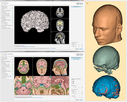
Scalp / cortex reconstruction
Segmentation and individual head models (FEM)
Following the automatic inhomogeneity correction the scalp and cortex of the subject are automatically segmented. The reconstruction of the scalp surface is done by segmenting the subject’s head from the background using a level set algorithm. Optimal results are achieved in an iterative process. Cortex segmentation is based on a white matter classification. The result is carefully smoothed and inflated to generate the cortical surface. This surface can be used as an approximate source space for cortical source imaging.
A full cortex inflation is also available as an optional step. If an MRI with cortex inflation is co-registered with a sensor cloud, the inflated cortex is automatically available in the BESA Research Source Analysis window for the visualization of cortical methods like Cortical CLARA, Cortical Loreta, or Minimum Norm estimate (BESA Research version 6.1 and higher; see also Source Analysis and Imaging).
In an optional step the individual (FEM) head model can be generated based on the segmentation results (see Wolters et al. 2007) and surface meshes of the 4 tissue layers (skin, scalp, CSF and brain) are extracted. The individual FEM head models can be used for EEG or MEG source analysis.
Automatic individual head (FEM) generation
- BESA MRI segmentation requires a few minutes of the users attention for preparing each subjects data set, thereafter it performs an automatic segmentation of the MRI data and generates a 4-layered FEM model (scalp, skull, CSF and brain).
- BESA MRI provides the possibility of registering T1 weighted MRIs with the respective T2 MRIs. This way the automatic segmentation can differentiate more accurately between tissues.
- The figure shows an example of cortex segmentation obtained by BESA MRI, as well as the respective FEM models for the scalp (orange), skull (green), CSF (blue) and brain (red). BESA MRI uses this 4-layered FEM model co-registered with EEG electrodes and / or head surface points to compute the lead fields which can be exported to BESA Research for source analysis.
Recent Comments