- New online workshop scheduled
See our workshop post for more information
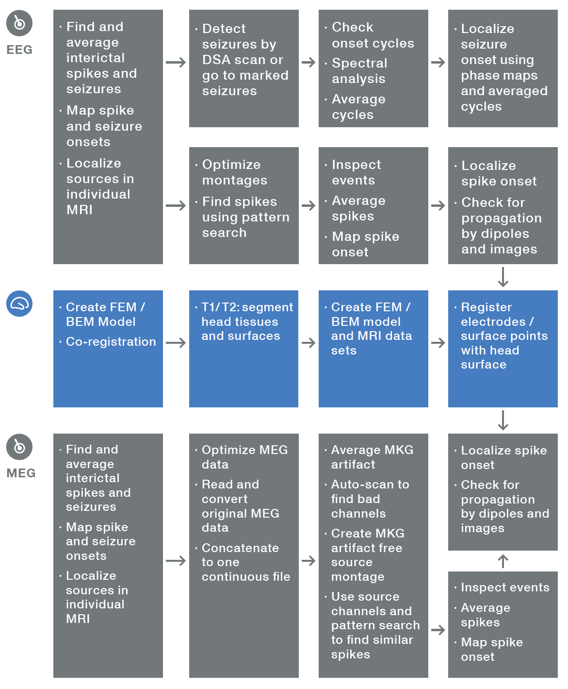
BESA pipeline
Analyze interictal spikes and seizure onset in EEG and MEG Research
The BESA pipeline is the seamless combination of BESA Research 7.1 and BESA MRI 3.0. Introducing the perfect aide in evaluating epileptiform interictal and ictal activity in EEG and MEG.
A comprehensive tutorial explaining step by step how to use BESA MRI and BESA Research for an optimized analysis of interictal and ictal activity in EEG is available:
BESA Research 7.1 – Tutorial for analyzing interictal spikes and seizure onset
Interictal activity (spikes)
Use BESA Research for reviewing patient data. To make decisions easier, BESA Research supports the evaluation of interictal activity with 3D Mapping, source montages and filters optimized for evaluation of epilepsy data.
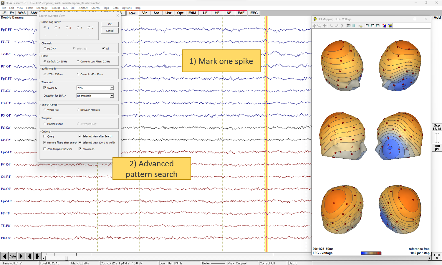
Spike Hunting: Find the first spike, let the automated search engine do the rest!
Just mark the first spike manually and then run the fully adjustable pattern search to find similar events quickly and reliably. Optionally, refine the search pattern using the auto-averaged spikes.
Average spikes for discrete or distributed source analysis. Analyze spike onset and check for propagation. Finally perform a source analysis using the averaged data to analyze e.g. the
onset phase.
Use BESA MRI for the segmentation of MR data and for a co-registration with the EEG. You can calculate an individual head model, and use it in the source analysis of BESA Research to obtain superior localization results.
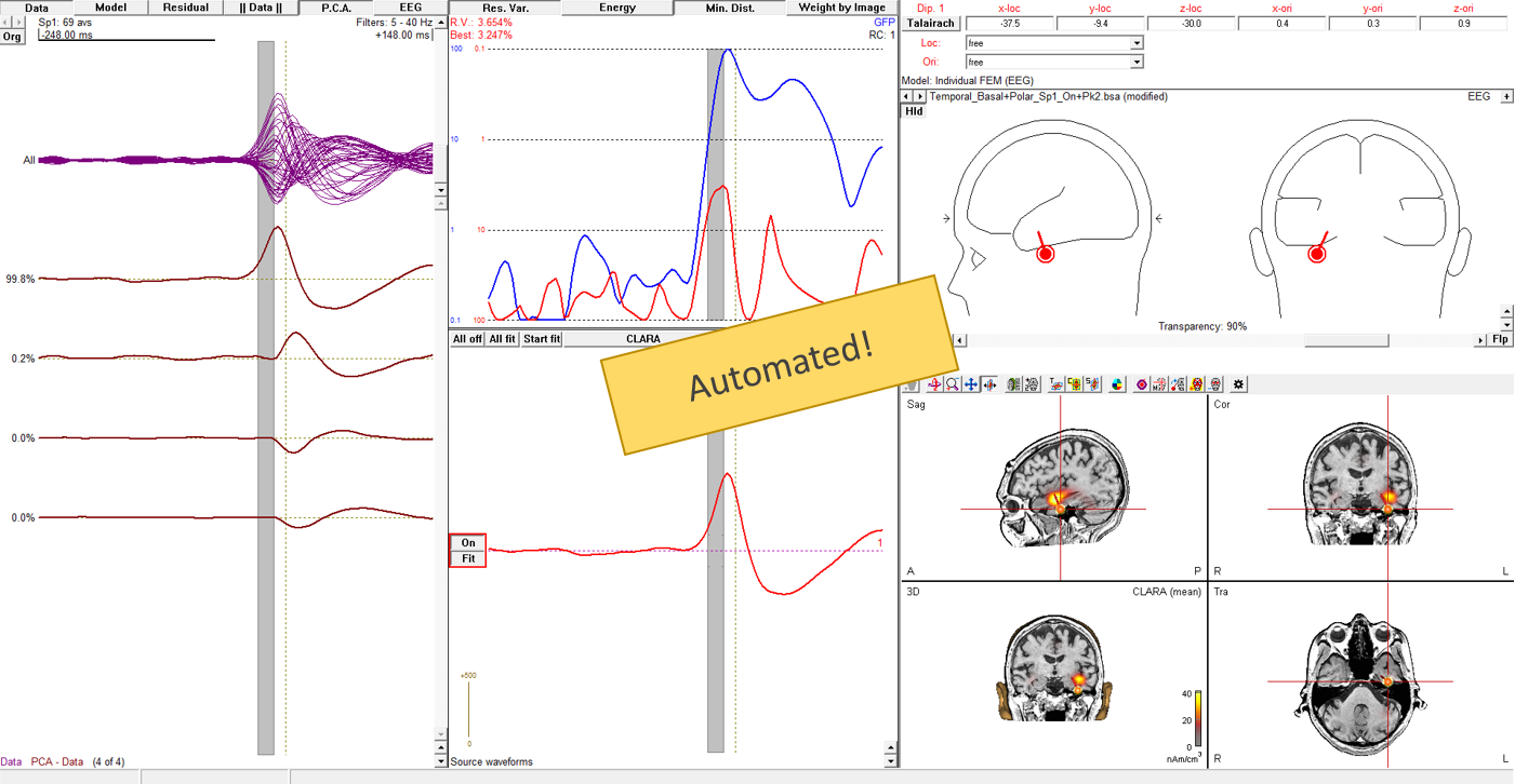
Source analysis: Get from a spike to its source in 5 mouse clicks!
Interictal spike analysis in MEG
This video demonstrates how to analyse interictal spikes in MEG, using pattern search and dipole source localization.
Ictal activity (spikes)
Use the customizable BESA Research DSA module to search for seizures and to analyze e.g. the onset phase. Use FFT and BESA Research’s FFT 3D phase maps to visualize and understand frequency (band)-specifi c activity and propagation.
Use BESA MRI 3.0 in the same way as for the interictal activity.
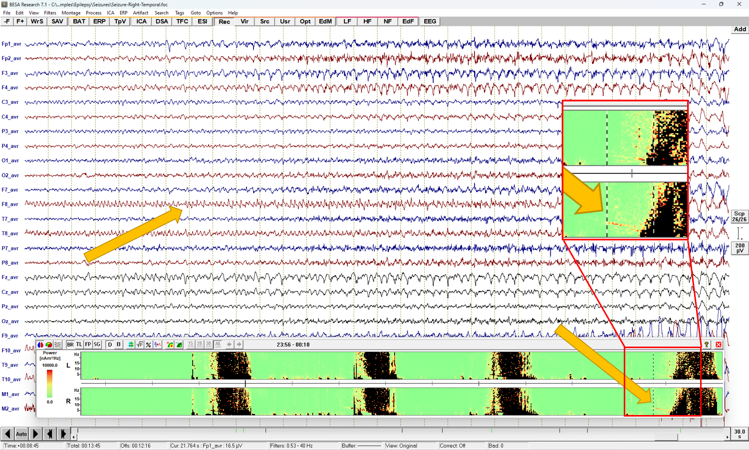
Chirp preceding seizure depicted by DSA in right hemisphere points to propagating right-temporal seizure
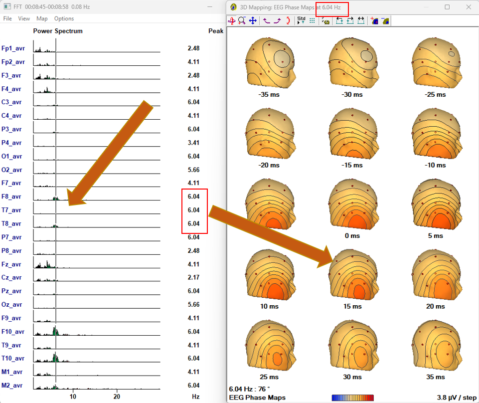
FFT phase maps show propagation to anterior temporal lobe
Recent Comments