This site presents the products of BESA GmbH, the leading innovators in digital EEG and MEG software for research and clinical applications.
- New online workshop scheduled
See our workshop post for more information
New features in BESA Research 7.1
BESA Research 7.1 provides the ideal combination of state-of-the-art research tools and proven diagnostic workflows in M/EEG analysis of human brain signals. Whether you are aiming for unlocking mechanisms of brain connectivity and function, or for enhanced diagnostic use on epileptiform data, BESA Research 7.1 will help you to achieve your targets reliably and with more pathways than ever before. This further expands on the set of new features that was introduced in the previous version BESA Research 7.0.
Here are some particular highlights of this exciting release:
Source analysis and source imaging
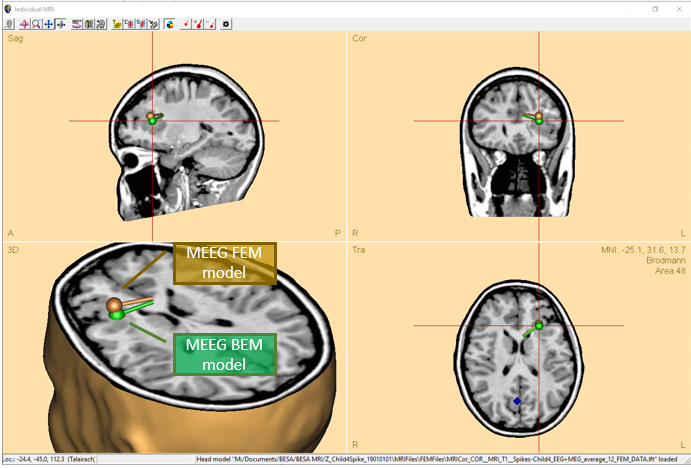
- For the first time, Boundary element method (BEM) and Finite element method (FEM) models can be directly compared on M/EEG data with just a couple of mouse clicks! After calculating the models automatically in BESA MRI, the models are automatically loaded into the Source analysis module and can be exchanged by two clicks in the head model section of the module.
- MEG and EEG can now be combined into a single data set for simultaneous source modeling of the two modalities. This is available for many source analysis and source imaging methods.
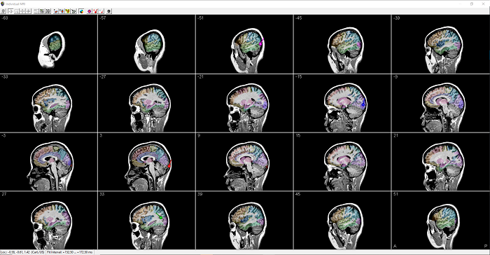
- MRI display now offers multi-slice view in any orientation for an easier review of solutions in the individual anatomy.
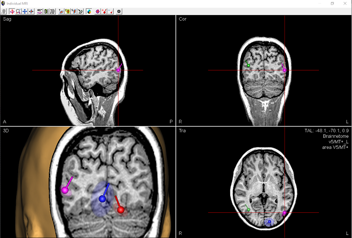
- Confidence limits are calculated for discrete source solutions, and displayed in co-registered MRI images.
- The Bayesian source imaging method SESAME (for an overview, see the Frontiers article) was improved to enhance robustness, as well as speed of computation and convergence. For this purpose, hyper-priors were introduced (cf. https://arxiv.org/abs/2006.04141) and parallel computing was optimized for this method.
- The full noise covariance matrix computed from individual trials can now be used in computation of minimum norm estimates.
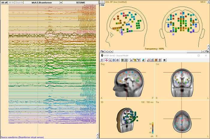
- Calculation of beamformer virtual sensor montages based on atlas regions is now supported.
- Two new brain atlases were added: Yeo7 and Yeo17 (https://doi.org/10.1152/jn.00338.2011).
- Montreal Neurological Institute (MNI) coordinates can now be used in the Source Analysis window.
- The baseline interval definition now features an automatic alert if it interferes with signal of interest.
- Ready-made color schemes for publication purposes are now available.
- Labels of discrete sources can be displayed in the 3D window.
Data review and pre-processing
- Atlas-based source montages: Pre-computed atlas-based source montages are now available from the menu entry Montage/Source/Atlas montages as well as under the Src button in the control ribbon.
- Parallel computing is used for speed-up of many time-consuming tasks.
- Smoother and faster plotting of waveforms eases review of high-density M/EEG data.
- For long-term EEG monitoring, an event histogram shows distribution of interictal spike events over the day.
- New data readers for XDF, Neuroscan CURRY 8 and CURRY 9 formats, and Dark Horse Neuro are available.
- OPM data by Cerca Magnetics can be read including sub-space projection for superior signal quality.
The full list of new features, improvements, and bugfixes is available here.
Download BESA Research 7.1 here.
Recent Comments