This site presents the products of BESA GmbH, the leading innovators in digital EEG and MEG software for research and clinical applications.
- First workshops of 2026 scheduled
Please visit our Workshops page for more information
Integration with MRI and fMRI
Integration with MRI and fMRI
For easy source analysis using individual MRI data, BESA Research provides an interactive interface to BESA MRI. EEG / MEG data can be coregistered with individual MRI data using individually digitized electrode positions or standard electrodes.
Coregistration and FEM model generation in BESA MRI
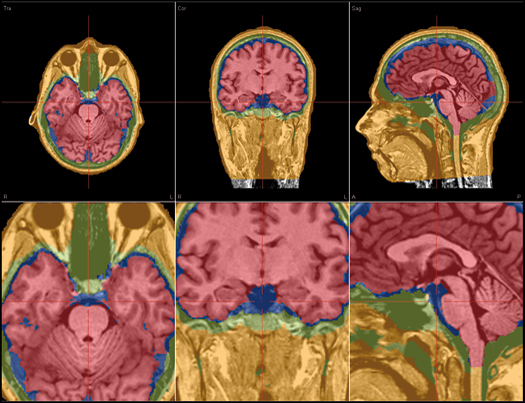
BESA MRI
- Integrated workflow for all user-interactions
- Automatic preprocessing with inhomogeneity correction
- Automatic reconstruction of scalp and cortex
- Automatic generation of individual 4-layer FEM model (scalp, skull, CSF and brain)
- Co-registration using digitized electrodes / headshape points or 10-10 / 10-20 standard electrodes
- Generation of FEM leadfields for individual head models co-registered with EEG electrodes and / or head surface points
- Overlay of source reconstructions on the individual MRI in BESA Research
- Separate license required for BESA MRI program
BESA Research also provides an interactive link to Rainer Goebel’s BrainVoyager™ (BV) program. The bidirectional connection of the two programs allows for source seeding from fMRI clusters with one mouse click.
BrainVoyager™
- Direct and easy interactive user interface of BESA Research with BV
- Analysis of individual MRI and fMRI data in BV
- Visualization and processing of individual MRI and fMRI
- Automated rendering of scalp and cortical surfaces
- Expansion and flattening of the cortical surface
- Minimum norm current image based on individual gray/white matter boundary
- Seeding of sources into BESA Research from anatomical 2D or 3D MR images or from fMRI BOLD clusters in BV via interactive link
- Overlapped display of fMRI and EEG / MEG sources in BV
- Separate license required for the BrainVoyager™ program
Recent Comments